Post-tuberculosis bronchial stenosis and bronchiectasis
Estenosis bronquial y bronquiectasias postuberculosis

This work is licensed under a Creative Commons Attribution-NonCommercial-ShareAlike 4.0 International License.
Ninguna publicación, nacional o extranjera, podrá reproducir ni traducir sus artículos ni sus resúmenes sin previa autorización escrita del editor; sin embargo los usuarios pueden descargar la información contenida en ella, pero deben darle atribución o reconocimiento de propiedad intelectual, deben usarlo tal como está, sin derivación alguna.
Show authors biography


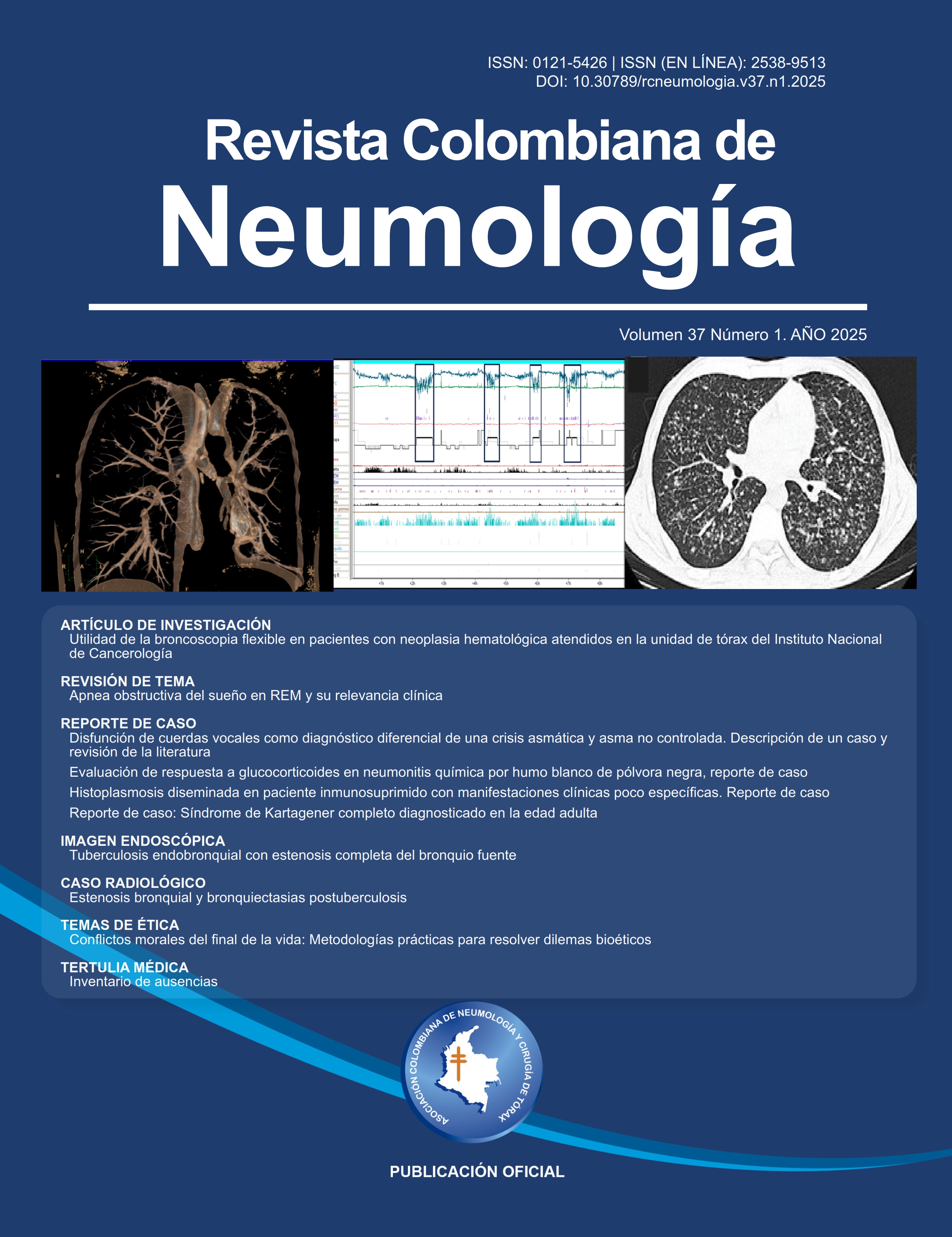
Pulmonary tuberculosis (TB) remains a common disease, especially in developing or underdeveloped countries. We present the case of a young female patient who was diagnosed and treated for pulmonary TB, with bacteriological improvement but with persistent respiratory symptoms, especially wheezing located in the left hemithorax.
A chest CT scan showed an obstruction of the left main bronchus, atelectasis, and bronchiectasis in the left upper and lower lobes.
The diverse types of sequelae secondary to pulmonary TB are presented, and the diagnostic means to differentiate relapses of the disease from the sequelae of TB and the possible treatments according to the type of lesion are discussed.
Article visits 128 | PDF visits 226
Downloads
- World Health Organization. Global tuberculosis report 2024 [Internet]. Geneva: WHO; 2024. [citado el 9 de marzo de 2025]. Disponible en: https://www.who.int/teams/global-tuberculosis-programme/tb-reports/global-tuberculosis-report-2024
- Nightingale R, Carlin F, Meghji J, McMullen K, Evans D, van der Zalm MM, et al. Post-TB health and wellbeing. Int J Tuberc Lung Dis. 2023;27(4):248-83. doi: http://dx.doi.org/10.5588/ijtld.22.0514 DOI: https://doi.org/10.5588/ijtld.22.0514
- Sehgal IS, Dhooria S, Muthu V, Salzer HJF, Agarwal R. Burden, clinical features, and outcomes of post-tuberculosis chronic obstructive lung diseases. Curr Opin Pulm Med. 2024;30(2):156-66. doi: http://dx.doi.org/10.1097/MCP.0000000000001026 DOI: https://doi.org/10.1097/MCP.0000000000001026
- Mbelele PM, Sabiiti W, Heysell SK, Sauli E, Mpolya EA, Mfinanga S, et al. Use of a molecular bacterial load assay to distinguish between active TB and post-TB lung disease. Int J Tuberc Lung Dis. 2022;26(3):276-8. doi: http://dx.doi.org/10.5588/ijtld.21.0459 DOI: https://doi.org/10.5588/ijtld.21.0459
- Migliori GB, Marx FM, Ambrosino N, Zampogna E, Schaaf HS, van der Zalm MM, et al. Clinical standards for the assessment, management and rehabilitation of post-TB lung disease. Int J Tuberc Lung Dis. 2021;25(10):797-813. doi: http://dx.doi.org/10.5588/ijtld.21.0425 DOI: https://doi.org/10.5588/ijtld.21.0425
- Taylor J, Bastos ML, Lachapelle-Chisholm S, Mayo NE, Johnston J, Menzies D. Residual respiratory disability after successful treatment of pulmonary tuberculosis: a systematic review and meta-analysis. EClinicalMedicine [Internet]. 2023;59(101979):101979. doi: http://dx.doi.org/10.1016/j.eclinm.2023.101979 DOI: https://doi.org/10.1016/j.eclinm.2023.101979
- Seo W, Kim HW, Kim JS, Min J. Long term management of people with post-tuberculosis lung disease. Korean J Intern Med. 2024;39(1):7-24. doi: http://dx.doi.org/10.3904/kjim.2023.395 DOI: https://doi.org/10.3904/kjim.2023.395
- Migliori GB, Blackbourn HD. Post-TB lung disease - the rationale for a Clinical Statement. Int J Tuberc Lung Dis. 2023;27(4):243-4. doi: http://dx.doi.org/10.5588/ijtld.22.0674 DOI: https://doi.org/10.5588/ijtld.22.0674
- Kim H. Rigid bronchoscopy for post-tuberculosis tracheobronchial stenosis. Tuberc Respir Dis (Seoul). 2023;86(4):245-50. doi: http://dx.doi.org/10.4046/trd.2023.0017 DOI: https://doi.org/10.4046/trd.2023.0017
- Choe KO, Jeong HJ, Sohn HY. Tuberculous bronchial stenosis: CT findings in 28 cases. AJR Am J Roentgenol [Internet]. 1990;155(5):971-6. doi: http://dx.doi.org/10.2214/ajr.155.5.2120966 DOI: https://doi.org/10.2214/ajr.155.5.2120966
- Deshpande SS, Joshi AR, Shah A. Aftermath of pulmonary tuberculosis: computed tomography assessment. Pol J Radiol. 2020;85(1):e144-54. doi: http://dx.doi.org/10.5114/pjr.2020.93714 DOI: https://doi.org/10.5114/pjr.2020.93714
- Caminero JA. A Tuberculosis Guide for Specialist Physicians. Paris-France: International Union Against Tuberculosis and Lung Disease (IUATLD); 2004.
- Karamchand S, Williams M, Naidoo P, Decloedt E, Allwood B. Post-tuberculous lung disease: should we be using Theophylline? J Thorac Dis. 2021;13(2):1230-8. doi: http://dx.doi.org/10.21037/jtd-20-1298 DOI: https://doi.org/10.21037/jtd-20-1298
Information
Journal history
Keywords
Directories and indexes
![]()


![]()
![]()


Support + Info Here
